

3D models of bones, joints, muscles, organs, and systems are available for check out. Pick up at the Circulation Desk on the 1st Floor of Lied Library.
Checkout policy
- Models are loaned for 4 hours.
- Late fees are $10 and accrue every hour that the model is late.
- The loaned period cannot be extended or renewed.
- Students must wait 30 minutes to check out the same model again.
- An alternative model of the same kind can be checked out if one is available.
- Models must stay inside Lied Library at all times.
- One pointer is checked out with each model to avoid the use of writing utensils as pointers.
Bones and Joints

Appendicular Bones Set
Disarticulated set of appendicular bones.
12 parts: wired hand, wired foot, scapula, clavicle, humerus, ulna, radius, femur, tibia, fibula, pelvis, and patella.
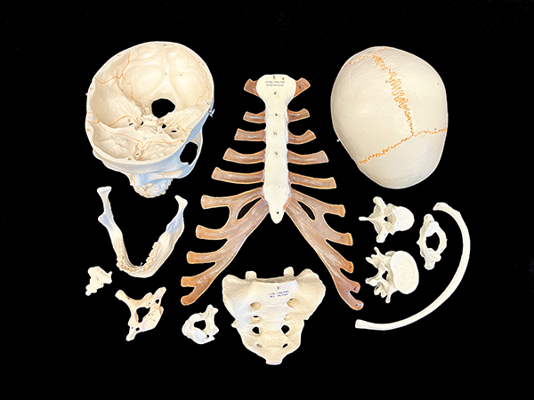
Axial Bones Set
Disarticulated set of axial bones
12 parts: sacrum, coccyx, mandible, rib, skull cap, base of skull, sternum, lumbar vertebra, thoracic vertebra, cervical vertebra, atlas, and axis.
Bone Pelvis
Female Pelvis: Contains hip bone, sacrum with coccyx and 2 lumbar vertebrae. Moveable Symphysis.
Male Pelvis: Contains hip bone, sacrum with coccyx and 2 lumbar vertebrae.
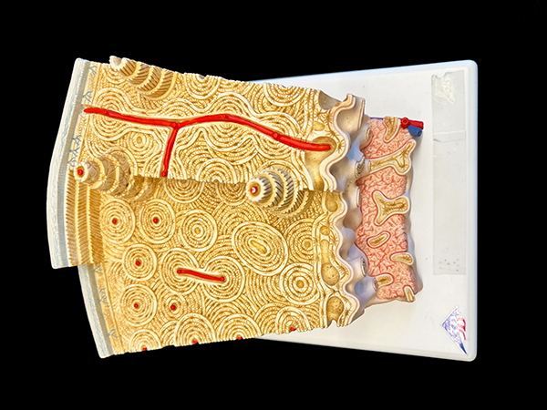
Bone Structure
Detailed histological structure of the bone, enlarged 80 times.
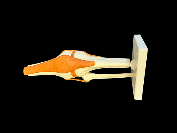
Flexible Knee
Life-size functional joint model, shows the anatomy and mechanics of the knee joint.
One part consists of a portion of femur, tibia, and portion of fibula, meniscus, patella, quadriceps tendon, and joint ligaments including the ACL and PCL.
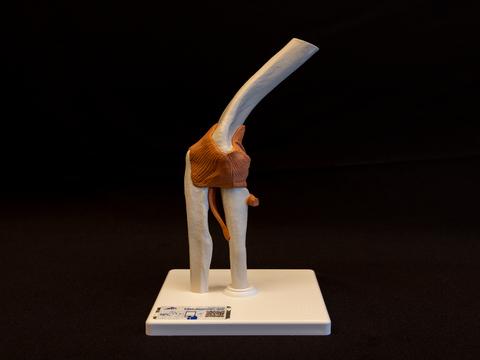
Human Elbow Joint
Model contains a portion of humerus, complete ulna and radius along with joint ligaments. Model is flexible and displays abduction, anteversion, retroversion, and internal/external rotation.
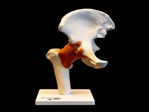
Human Hip Joint
Model contains portions of femur, hip bone, and joint ligaments. Model is flexible and displays abduction, anteversion, retroversion, and internal/external rotation.
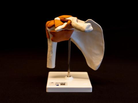
Human Shoulder Joint
Model contains scapula, clavicle, portion of humerus and joint ligaments. Model is flexible and displays abduction, anteversion, retroversion, and internal/external rotation.
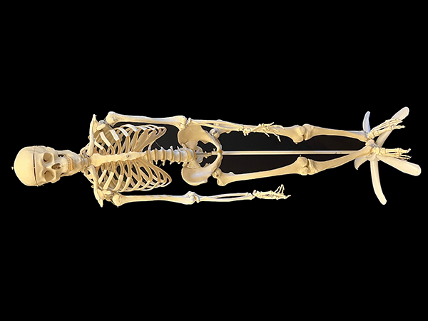
Human Skeleton, "Mandy"
Full human skeleton model, assembled.
Note: Mandy the Skeleton is listed as Stan in the library system.
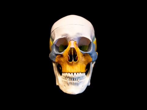
Multicolored Human Skull
Different colors are portrayed on the skull to help study the position and relationship of skull structures. Skull cap is removeable.
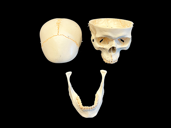
Numbered Classic Skull
3 parts: skull cap, base of skull, and mandible.
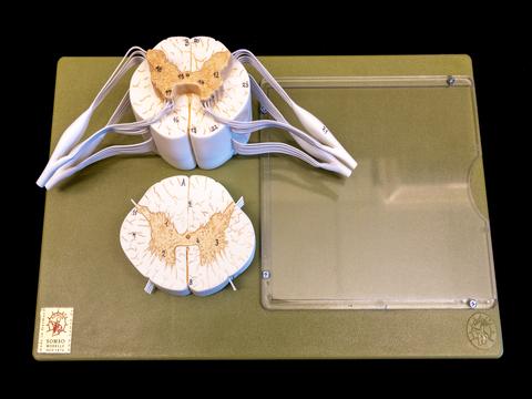
Spinal Cord Histology
Portion of spinal cord which shows spinal nerves at approximately 5x life size. Cross section is approximately 10x life size.
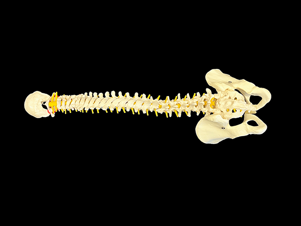
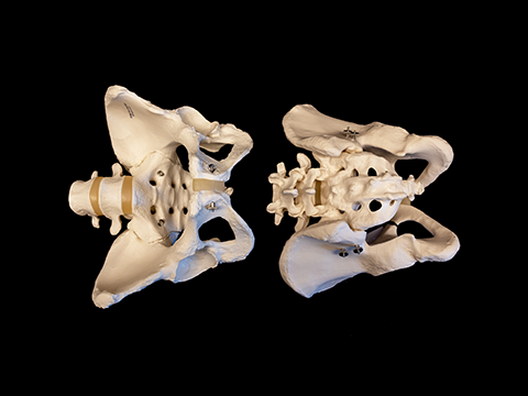
Vertebral Column and Pelvis
Female Pelvis
Full pelvis and occipital plate, fully flexible mounting throughout spine, L3-L4 disc prolapsed on spine, spinal nerve exits, and cervical vertebral artery.
Muscles
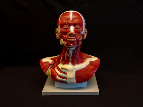
Human Head and Neck Model
Displays muscular and vascular structures in the head and neck, approximately life size.
Head Model
Outer, superficial, and internal (median section) of structures of the head and neck.
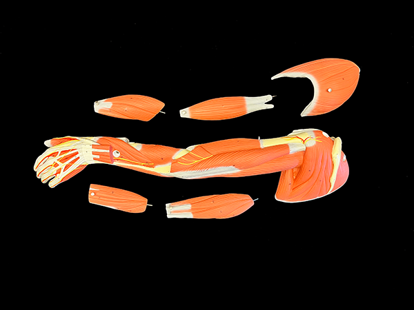
Muscled Arm
Superficial and deeper muscles including tendons, vessels, nerves, and bone components of the left arm and shoulder.
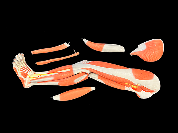
Muscular Leg
Superficial and deeper muscles including tendons, vessels, nerves, and bone components of the left leg and foot.
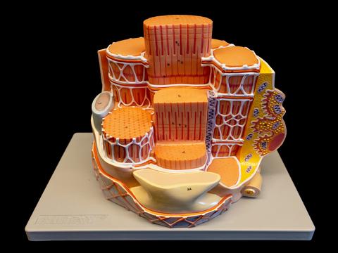
Skeletal Muscle Fiber Model
Approximately 10,000 times life size. Model displays microstructure of skeletal muscle fiber. Model includes neuromuscular junction, sarcoplasmic reticulum, T tubules, and myofibrils.
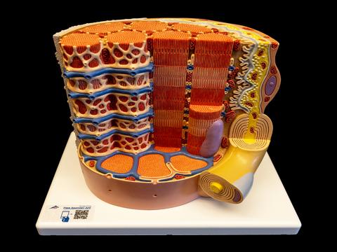
Skeletal Muscle Histology Model
Approximately 10,000 times life size. Model displays a section of the skeletal muscle fiber in association with the neuromuscular end plate.
Muscular Torso
Deep and superficial muscles.
25 parts total: chest/abdominal wall, 2 part larynx, 2 lungs, diaphragm, 2-part stomach, liver with gallbladder, intestinal tract with appendix, front half kidney, half urinary bladder, and 4 muscles.
Organs and Systems
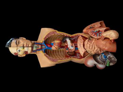
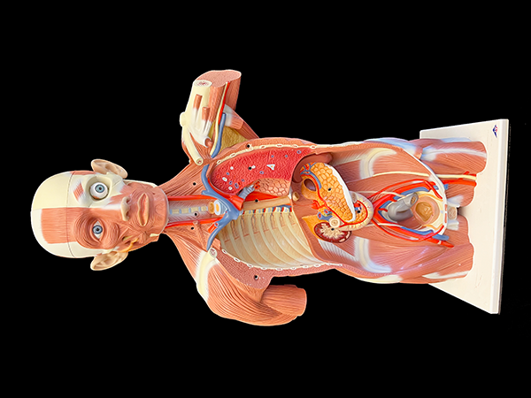
Anatomical Torso, "Clark"
18 parts total: head (2 parts), brain (1 half), lungs (4 parts), heart, esophagus, descending aorta, diaphragm, stomach, duodenum with pancreas & spleen, intestines, kidney, liver with gallbladder, bladder (2 parts)
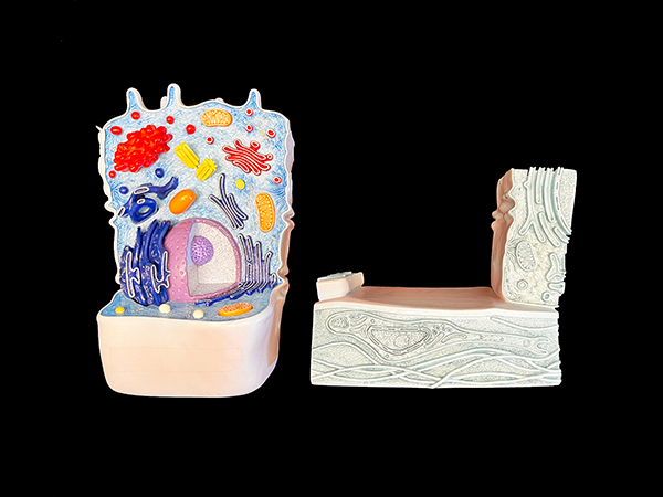
Animal Cell
Shows the form and structure of a typical animal cell as viewed by an electron microscope.
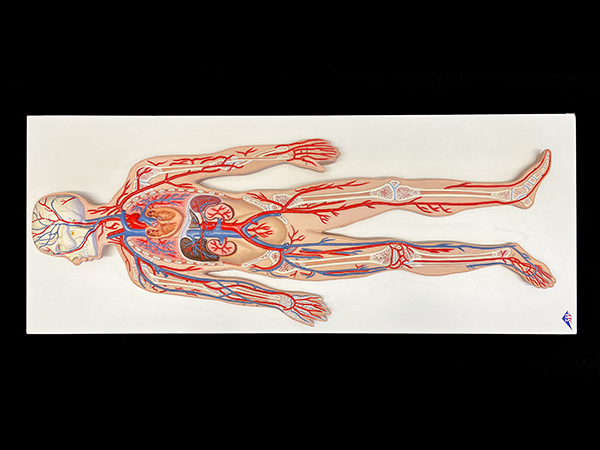
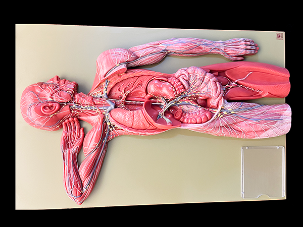
Blood Circulatory System
Half size relief model of the human circulatory system. Shows detail of the arterial/venous system, heart, lung, liver, spleen, kidneys, and partial skeleton.
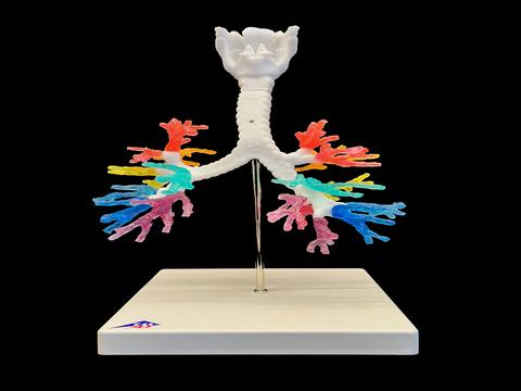
CT Bronchial Tree Model
Model displays a larynx with hyoid bone, epiglottis, trachea, and lobar bronchi.
Digestive System
Life-size digestive system model that demonstrates the entire digestive system in graphic relief.
Features nose, mouth cavity and pharynx, esophagus, GI tract, liver with gallbladder, pancreas, and spleen.
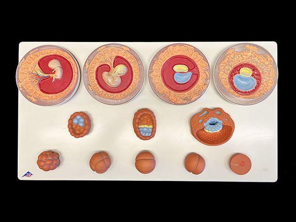
Embryonic Development Stages
Embryonic development model in 12 stages. The model represents the development of the human germ cells from fertilization until the end of the 2nd month of pregnancy in the 12 stages.
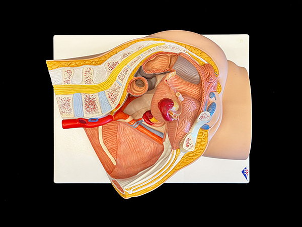
Female Pelvis
Two-part median section of female pelvis.
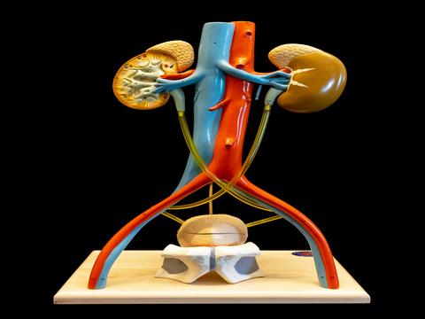
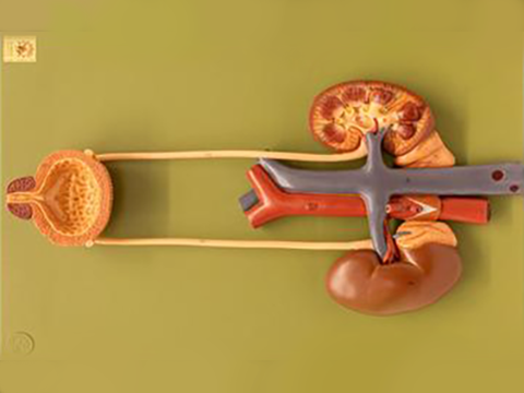
Free-Standing Urinary System
Model displays kidneys, adrenal glands, abdominal aorta/branches, inferior vena cava/branches, iliac vessels, ureter, upper half of bladder and prostate.
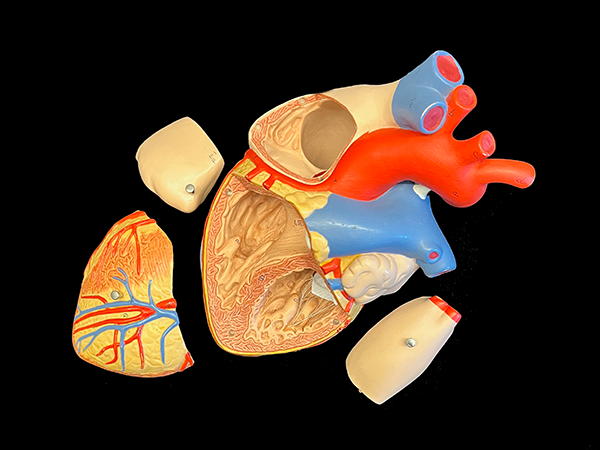
Heart Model
Two times life size heart model. Atrium walls and the front of the heart wall are removable to reveal a professionally detailed and realistic heart.
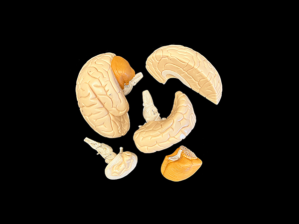
Human Brain
Life-size and anatomically correct
3 different models: 2-part, 5-part, and 4-part.
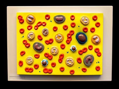
Human Blood Cell Type Model
Model displays erythrocytes, platelets, and 5 types of leukocytes. Model also shows leukocytes characteristic nuclei.
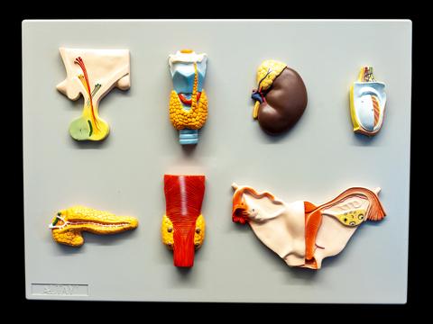
Human Endocrine Organs Model
Approximately life-size model featuring the pituitary, adrenal, thyroid, and parathyroid glands, along with a testis and a dissected ovary with fallopian tubes.
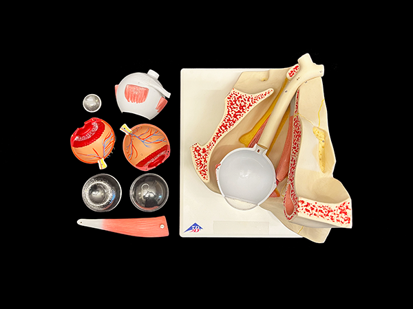
Human Eye
Three times life size anatomical human eye model shows the optic nerve in its natural position in the bony orbit of the eye (floor and medial wall).
Can be dissected into both halves of sclera with cornea and eye muscle attachments, both halves of the choroid with iris and retina, eye lens, and vitreous humor.
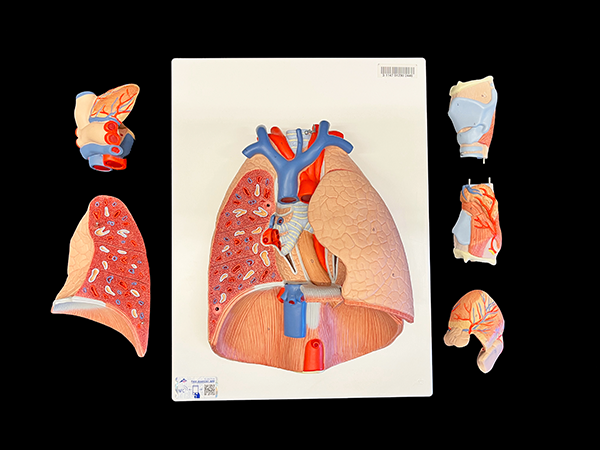
Human Lung with Larynx
Contains the following removable parts: 2-part larynx, trachea with bronchial tree, 2-part heart, subclavian artery and vein, vena cava, aorta, pulmonary artery, esophagus, 2-part lung, and diaphragm.
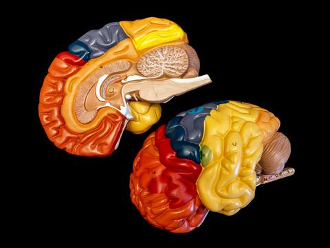
Human Regional Brain
Approximately life size color coded two part brain. The color coding assists in identifying the primary functional areas within the cerebral cortex.
Human Urinary System
Approximately life size model of human urinary system in color. The colors assist in better identifying the system parts.
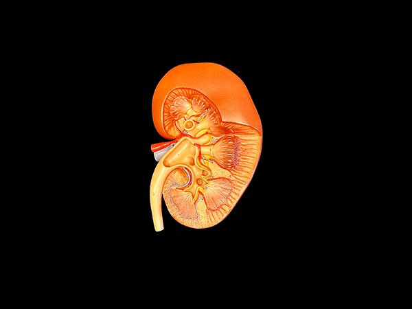
Kidney Section
Three times life size kidney section model depicts a longitudinal section of the right kidney.
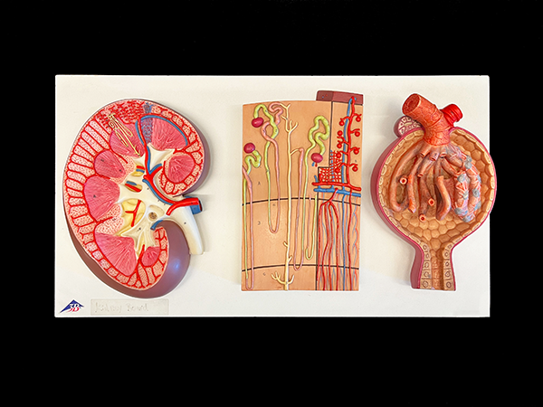
Kidney Section, Nephron, and Renal Corpuscle
3 kidney models mounted on one baseboard.
Three times life-size longitudinal section of right kidney. 120 times full-size kidney nephron. 700 times life-size opened Malpighian corpuscle.
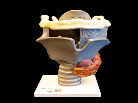
Larynx Model
Model includes moveable epiglottis, vocal cords, and arytenoid cartilage. Model also displays hyoid bone, cricoid cartilage, thyroid cartilage, thyroid, and parathyroid glands.
Lymphatic System in the Human Body
Lymphatic vessels, ducts, and lymph nodes.
Two thirds life size, 1 part relief model depicts the human lymphatic system.
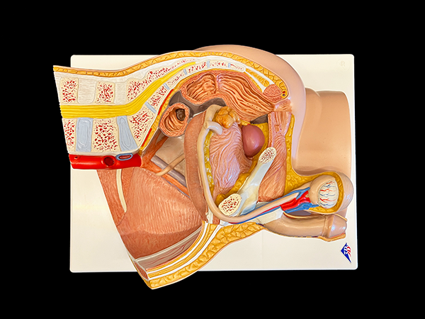
Male Pelvis
2-part, median section of male pelvis which presents parts of the abdominal and dorsal muscular system of the human male with lumbar vertebrae and partial kidney with removable bladder and reproductive organs.
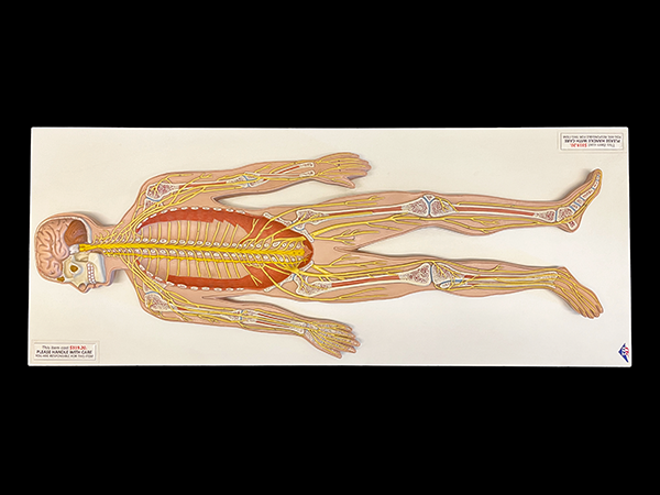
Nervous System
Half size nervous system relief model shows a schematic representation of the central and peripheral nervous system.
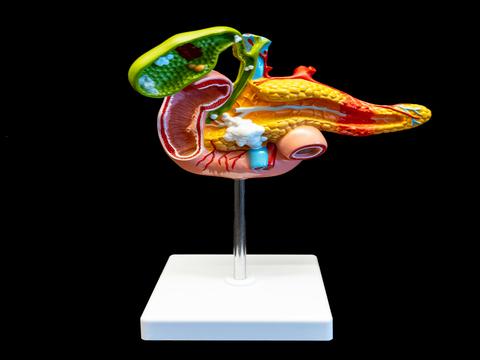
Pathology of the Human Pancreas, Duodenum, and Gallbladder
Partially opened model shows stones in multiple locations, including cholecystitis, polyposis, and carcinoma. Highlights key diseases of the gallbladder, pancreas, and duodenum—such as pancreatitis in the pancreas tail, cancer in the pancreas head, and a duodenal ulcer.
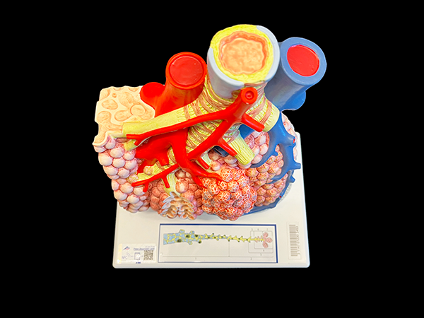
Pulmonary Lobule with Surrounding Vessels
1-part, 130 times magnification of external pulmonary lobule.
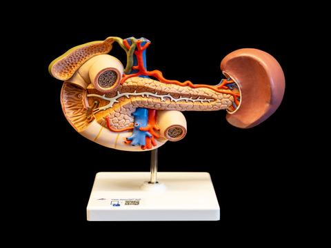
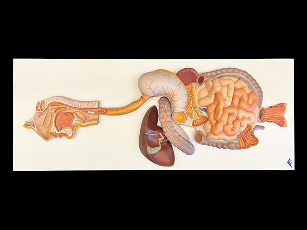
Rear Organs of Upper Abdomen
Model displays a partially opened duodenum, an opened gallbladder/bile ducts/ pancreas/spleen and surrounding vessels in natural size.
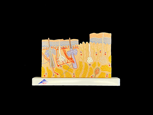
Small Skin Model
Human skin section model, 40 times life size.
Features detail with hair follicles, sebaceous glands, sweat glands, receptor nerves, and vessels.
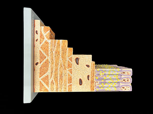
Structure of Stomach Wall
Microstructure within a section of the human stomach wall.
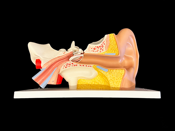
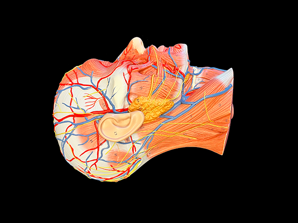
Vestibulocochlear Organ (ear)
The human ear model represents the outer, middle, and inner ear.
Removable eardrum with hammer, anvil, and stirrup as well as labyrinth with cochlea and auditory/balance nerve.